Physiology and Behaviour
Explain one study related to localization of function in the brain (for example, Wernicke, Broca, Gazzaniga and Sperry).
|
Broca's Aphasia
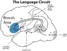
Name: Broca's Aphasia (non-fluent aphasia) Researcher: Paul Broca Date: 1861 Definition: Speech output is severely reduced and is limited mainly to short utterances of less than four words. A person with Broca's aphasia can understand speech but have difficulties producing it. They are able to read, but are limited in writing. Vocabulary access is limited and the formation of sounds by persons with Broca's aphasia is often clumsy. Aim: Broca noticed some patients had problems with speech production and he wanted to find out if their impediment was related to the brain. Method: Broca dissected the brains of the patients to study damaged areas. Results: He found that there was damaged area in the left frontal lobe of the patients. Conclusion: Broca's area is localised for some aspects of speech production but Broca's aphasia can be a combination of various damages to brain areas beyond Broca's area. Evaluation:
|
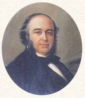
Paul Broca (1824 – 1880)
French surgeon, anatomist and anthropologist.
|
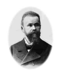
Carl Wernicke (1848-1905). German physician, psychiatrist and neuropathologist.
|
Wernicke's Aphasia
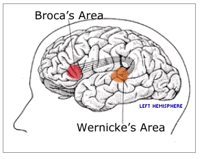
Name : Wernicke's Aphasia(fluent aphasia) Researcher: Carl Wernicke Date: 1874 Definition: In this form of aphasia the ability to grasp the meaning of spoken words is chiefly impaired, while the ease of producing connected speech is not much affected. Therefore Wernicke's aphasia is referred to as a 'fluent aphasia.' However, speech is far from normal. Sentences do not hang together and irrelevant words intrude-sometimes to the point of jargon, in severe cases. Reading and writing are often severely impaired. Aim: Wernicke wanted to prove that her patients could produce speech, but could not understand it. Method: 1.He used a reflex arc model (sensory and motor centers) to explain brain function. 2.He discovered the brain center for sensory aphasia or "Wernicke's aphasia". 3.He did careful descriptions of functional disturbances as well as pathological detail of the brain. 4.He developed a model of the brain based on differentiating brain centers, that works something like an information processing model to ascertain the location of brain lesions from symptoms and vice versa. Results: she first described the left posterior superior temporal gyrus and proved that patients could produce speech, but could not understand it. Conclusion: The research undertaken by Broca and Wernicke provides us with a clear understanding of some of the factors involved in language processing. Evaluation: Eggert, Gertrude H. (1977). Wernicke's works on aphasia: A sourcebook and review: Early sources in aphasia and related disorders, Volume 1. The Hague: Mouton Publishers. |
Using one or more examples, explain effects of neurotransmission on human behaviour (for example, the effect of noradrenaline on depression).
|
Seratonin
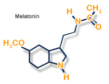
Serotonin Syndrome: When you have too much serotonin present in your body serotonin syndrome can occur. Sadock and Sadock report this syndrome occurs when plasma concentrations in the blood are raised to toxic levels. These high levels of serotonin will cause behaviors of restlessness and extreme agitation with the possibility of seizures. The Mayo Clinic reports that serotonin syndrome and the behavioral symptoms associated with it will return to normal once the serotonin levels are returned to normal. If you feel you need more information on this disorder consult with your doctor or pharmacist. Selective Serotonin Reuptake Inhibitors: Selective Serotonin Reuptake Inhibitors, SSRIs, are a line of drugs that inhibit the reuptake of serotonin by the brain, reports Sadock and Sadock. When serotonin reuptake it blocked the brain has more serotonin available to it. This enhances and increases the transmission of the neurotransmitter, serotonin which can affect your mood and behavior, according to the Mayo Clinic. These drugs aid in stabilizing behaviors associated with mental health disorders, such as depression and obsessive-compulsive disorders among others, as reported by Sadock and Sadock. Behavioral Disorders: Serotonin has been show to influence a variety of behavioral disorders and a first line of defense in therapeutic treatments are drugs that influenced serotonin levels in the brain; including obsessive compulsive disorder, panic disorder, social phobia, posttraumatic stress disorder, eating disorders, attention-deficit hyperactivity disorder and insomnia, as reported by Sadock and Sadock. In these disorders the serotonin levels appear to be contributing to negative behaviors because once the patient is starting on drugs that influence the levels of brain serotonin a reduction in their negative behaviors is usually seen, according to Sadock and Sadock. Suicide: It has been found that an increase of serotonin in the brain can cause a reduction in suicide overall, as reported by Sadock and Sadock. Despite the decrease, some patients who begin taking SSRI's increase their suicidal attempts with more success, according to Sadock and Sadock. They suggest the initial increase in serotonin due to the SSRI's could allow the patient enough rise out of depression to engage in the suicidal behavior; thus, patients with thoughts of suicide should be monitored during their first weeks on SSRIs. Once the patient has been on the SSRI's for a number of weeks their risk of suicide is decreased. Using one or more examples, explain functions of two hormones in human behaviour.
|
Discuss two effects of the environment on physiological processes (for example, effects of jet lag on bodily rhythms, effects of deprivation on neuroplasticity, effects of environmental stressors on reproductive mechanisms).
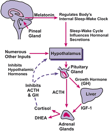
Jet Lag
Jet lag is a physiological condition caused by disturbance to the body’s natural circadian rhythm, or internal clock. This condition is largely caused by air travel across one or more time zones, from which its name is derived, but can also be caused by shift work or other factors.The effects or symptoms of jet lag generally last only a few days, but could last longer depending on the amount of disturbance the steps taken to combat them. Jet leg affects on Daytime sleepiness, Headaches, Insomnia Restless sleep, possibly with frequent awakening, Difficulty concentrating, Impaired judgment, Upset stomach or mild nausea. Example of the jet leg clearly proves that environment can really affect on the physiology. The second environmental factor is the effect of environment on reproduction.Consideration of environmental influences on human reproduction must include an investigation of the socioeconomic factors that play an important role in embryo-fetal development. Reproductive health is exquisitely sensitive to characteristics of an individual's environment —including physical, biological, behavioral, cultural and socioeconomic factors. The relative effects of these features may vary in different parts of the world or even within a country. Environmental changes such as global warming, increased radiation and exposure can really effect on human physiology.
Jet lag is a physiological condition caused by disturbance to the body’s natural circadian rhythm, or internal clock. This condition is largely caused by air travel across one or more time zones, from which its name is derived, but can also be caused by shift work or other factors.The effects or symptoms of jet lag generally last only a few days, but could last longer depending on the amount of disturbance the steps taken to combat them. Jet leg affects on Daytime sleepiness, Headaches, Insomnia Restless sleep, possibly with frequent awakening, Difficulty concentrating, Impaired judgment, Upset stomach or mild nausea. Example of the jet leg clearly proves that environment can really affect on the physiology. The second environmental factor is the effect of environment on reproduction.Consideration of environmental influences on human reproduction must include an investigation of the socioeconomic factors that play an important role in embryo-fetal development. Reproductive health is exquisitely sensitive to characteristics of an individual's environment —including physical, biological, behavioral, cultural and socioeconomic factors. The relative effects of these features may vary in different parts of the world or even within a country. Environmental changes such as global warming, increased radiation and exposure can really effect on human physiology.
Effect of environment of Reproduction
Examine one interaction between cognition and physiology in terms of behaviour (for example, agnosia, anosognosia, prosapagnosia, amnesia). Evaluate two relevant studies.
Prosapagnosia
Prosapagnosia is an inability to recognize faces including, perhaps, your own. There can be many reasons why a person may have problems with this, but it is not because they don't have good vision. It is a problem within the brain. Facial recognition occurs in the part of the brain known as the fusiform gyrus. It is located within the frontal region underneath part of the brain. If an injury occurs to this area, there is a brain tumour in the area or is removed, the person will have difficulty recognizing faces. Developmental prosopagnosia refers to an inability to recognize faces despite typical intellectual functioning, emotion and object recognition, and no evidence of brain injury. Very little is known about this disorder in children. We present a case study of a child, henceforth referred to as B, who reported extreme difficulty in recognizing faces.
Participant: B is a healthy 7 year old male with no history of neurological or behavioral disorder. His parents report that he is above-average in intelligence and very social. He has no history of visual impairment other than a deficit in face recognition.
Methods: We evaluated B's face recognition skills using a variety of measures for which we have been collecting normative data. We examined his ability to recognize faces, discriminate between faces, identify emotion, and recognize objects. A clinical neuropsychologist also evaluated B using a wide variety of measures. Finally, we employed a training program to improve B's ability to recognize the faces of familiar people in his life.
Results: On our test of face recognition, B answered correctly on 46.8% of trials (50% is chance); average performance of 8 year olds on this test is 80% . These results provide evidence of a severe deficit in face recognition. B showed no evidence of a similar deficit in the ability to discriminate faces, recognize objects, and identify emotion. The neuropsychological evaluation also found a deficit in face recognition despite intact object recognition, memory, emotion recognition, and above-average IQ. Finally, we successfully trained him to identify photographs of 30 familiar people; however, he used local characteristics of the photograph to recognize the faces.
Anosognosia
Impaired or lack awareness of illness - a neurological syndrome called anosognosia - is believed to be the single largest reason why individuals with schizophrenia and bipolar disorder do not take their medications. Caused by damage to the brain, it affects approximately 50% of individuals with schizophrenia and 40% of individuals with bipolar disorder. When taking medications, awareness of illness improves in some patients.
Still today anosognosia as a phenomenon attracts the attention of both clinicians and researchers. Patients affected by this syndrome deny an obvious neurological dysfunction caused by a defined damage in their brains. This lack of awareness of their impairment is the most interesting feature of this syndrome. In the neuropsychiatric literature case studies have played an important role, among them four contributions by Gabriel Anton (1858-1933) at the end of the 19th century. The phenomenon described by him in these case studies was later referred to as anosognosia by Josef François Babinski (1857-1932). Recognising his achievements in the description of this phenomenon one kind of anosognosia, namely cortical blindness, is still referred to in the scientific literature as the Anton-Syndrome (also Anton Symptom).
Using the recently discovered original files of 1895/96 our study substantiates one case described by Anton. The case in question is that of Juliane Hochriehser, a 69-year old dairymaid who showed anosognosia with cortical deafness due to bilateral lesion of the temporal lobes. Other cases of his are also included. The study concludes with an overview of the current state of research and the different approaches to this syndrome. Yet it is still not clear which areas and structures in the brain are responsible for the development of anosognosia. It may well be that dextral or bilateral damage of several areas of the brain plays a major role.
Discuss the use of brain imaging technologies (for example, CAT, PET, MRI) in investigating the relationship between biological factors and behaviour.
|
PET
Short form for position emission tomography. It is a technique that produces 3D image of functional processes in the body. It scans and monitors glucose metabolism in the brain and it can also record ongoing activity in the brain for example thinking. This technology is also very good for diagnosing tumors, changes in Alzheimer's and it can compare brain differences. PET technology is a great example of showing human behavior, because it can record the ongoing activity in the brain. 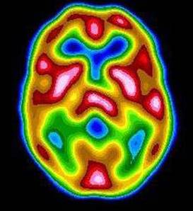
|
CAT
Stands for of Computerized Axial Tomography. CAT Scans can be used for various parts of the body, including the brain, spine, abdomen, chest, and pelvis. CAT scans are a combination of x-ray technologies and computer-imaging techniques. Therefore, CAT scans produced a more detailed picture than just an x-ray would. The machine itself is shaped somewhat like a donut, and the subject lays on a bench that slides through the center of the donut, where the machine captures the pictures of the body. This technology is also a great example of showing how our body works. 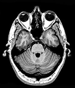
|
MRI
Magnetic resonance imaging (MRI) is a medical imaging technique used in radiology to visualize internal structures of the body in detail. MRI makes use of the property of nuclear magnetic resonance(NMR) to image nuclei of atoms inside the body. MRI provides good contrast between the different soft issues of the body, which makes it especially useful in imaging the brain, muscles, the heart, and cancers compared with other medical imaging techniques such as computed tomography(CT) or rays. Unlike CT scans or traditional X-rays, MRI does not use ionizing radiation. MRI scanner is a device in which the patient lies within a large, powerful magnet where the magnetic field is used to align the magnetization of some atomic nuclei in the body, and radio frequency fields to systematically alter the alignment of this magnetization.This causes the nuclei to produce a rotating magnetic field detectable by the scanner—and this information is recorded to construct an image of the scanned area of the body. |